


As a student of the Medical Art MSc course at the University of Dundee, I could not be more grateful for the access to cadaver dissection, and anatomical specimens throughout the course. My undergraduate degree was in Fine Art, where I specialized in classical figurative sculpture. As part of my undergraduate course, I completed several ecorche sculptures through study of a live model. It was not until coming to University of Dundee and having access to cadaver dissection while simultaneously completing a head and neck ecorche that I truly saw the value of dissection in the teaching and learning of anatomy. To be able to dissect facial structures in the lab, and replicating those same structures in clay in the studio, I believe, is the best way possible to learn anatomy. You experience the anatomy from both the exterior and interior, providing the artist with a full understanding.
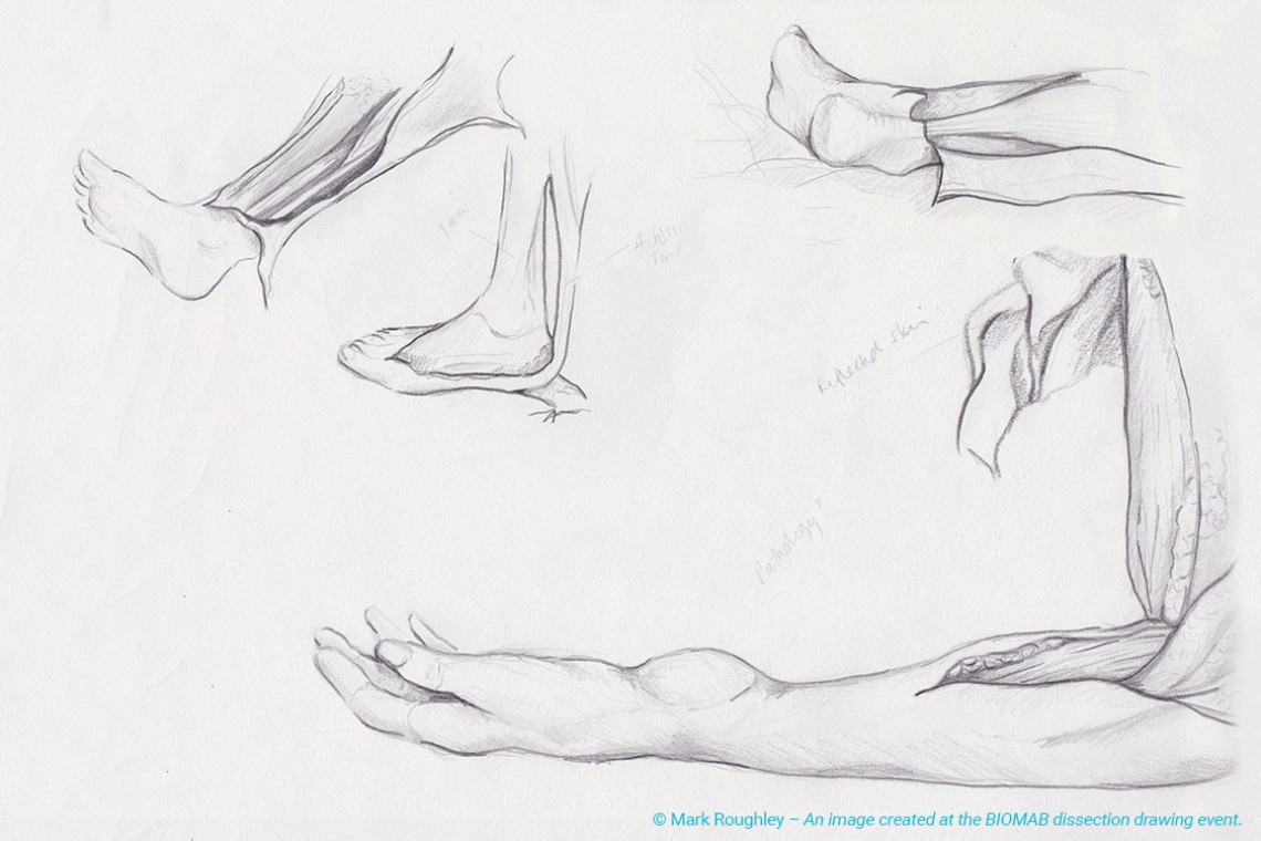
I was fortunate to be able to attend BIOMAB’s Dissection drawing days in April 2017. This provided for a very different but rewarding dissection experience in comparison to University of Dundee’s course. The cadavers at BIOMAB are fresh frozen, not embalmed, and ranged from a variety of animal and human specimens. Although University of Dundee’s cadavers are colourful with their Thiel embalming, nothing compares to the colours and “life” evident in the fresh frozen cadavers used by BIOMAB. In the making of anatomical art, we are often told to depict dissected specimens as if they are still alive and functioning in life. BIOMAB’s fresh frozen cadavers demonstrated to me exactly what to depict in my own art. The specimens were mostly human limbs and organs. Although these dissections were different from those at Dundee and did not correlate as they were different portions of the body, they did relate in respect to muscle form, function, origination and insertion. Nothing compares to the ability of actually feeling and following a muscle’s path through space on the human body, and being able to see its origin and insertion. It also is clearly evident what is muscle tissue, tendon, and ligament just by simply looking at the dissections and the form variations. These qualities were much more evident in BIOMAB’s dissections, rather than University of Dundee’s Thiele embalmed cadavers.
The instruction at BIOMAB was also superb, with demonstrators teaching in multiple languages simultaneously. Then artists also had the ability to take photographs of the specimens for individual use and reference, in which I will be using for my future ecorche sculpture studies.
As a student of MSc Forensic Art & Facial Identification at the University of Dundee I am focusing on the anatomy of the head and neck. At Dundee, I have had the opportunity to view and dissect the faces of full body Thiel-embalmed cadavers. Thiel-embalming is a method of preserving cadavers and is the process of choice by the University of Dundee. Preserving the cadavers in this way is beneficial because the elasticity is retained and therefore mimics a living-patient with a high degree of accuracy. We also have access to prosections where formalin embalming has been previously carried out, before the introduction of Thiel. This has allowed us to see one of the many benefits of using Thiel. The prosections are very firm to the touch in comparison to the Thiel-embalmed cadavers and therefore, in this way, the prosections are less realistic.
At the BIOMAB event, the cadavers were not Theil-embalmed, they were fresh frozen. Therefore, the colours were more vivid and also veins and arteries were easier to identify. Day one, we observed the students at the Faculty of Veterinary Medicine of Antwerp, Belgium, carrying out their work. It was interesting to be able to talk with the students and to observe. The animal anatomy I looked at was of full bodies.
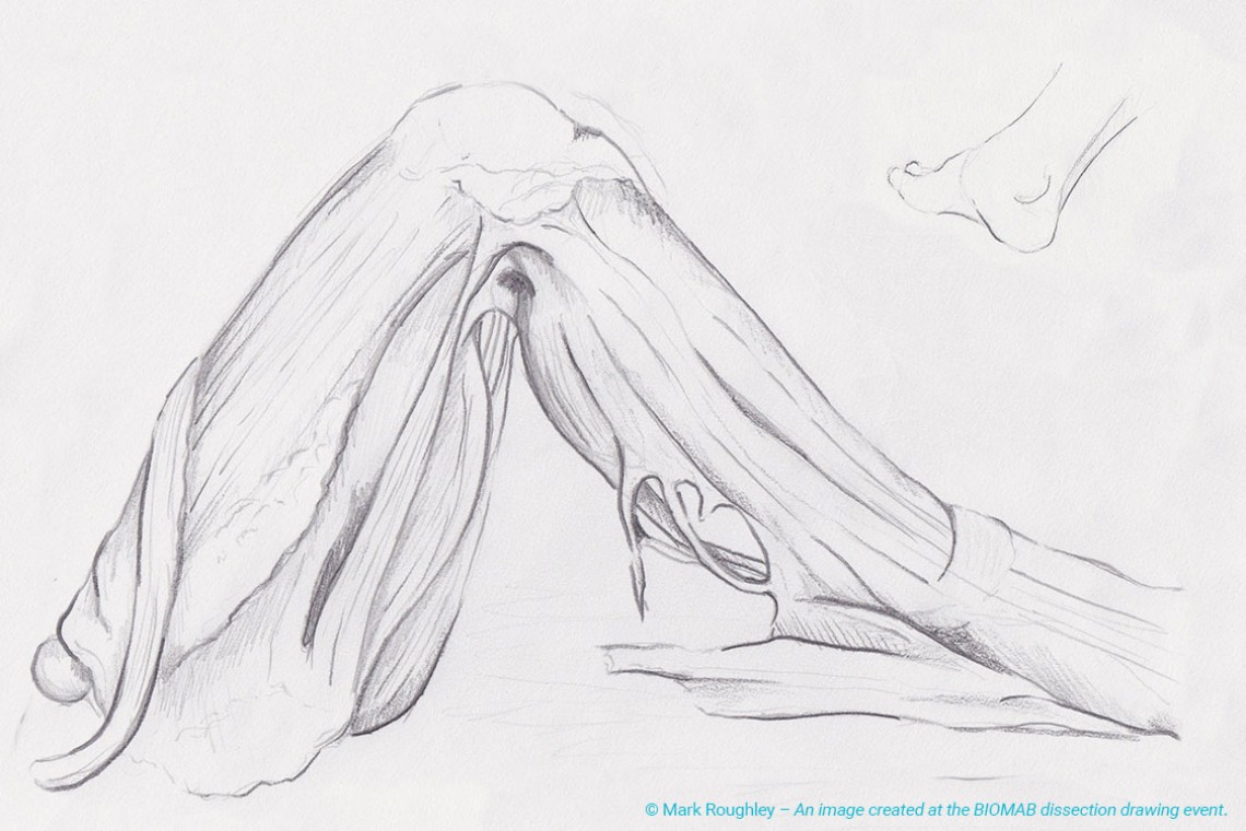
On the second day, at the Faculty of Medicine of Antwerp, fresh frozen human cadaver limbs were provided. We were grateful for the explanations and demonstrations carried out by the surgeons leading the dissections. Surrounding the dissections were other tables with models, skulls and in the same room, simultaneously, Ann Van de Velde carried out a facial reconstruction sculpture workshop. For me, in addition to the limbs, it would have been interesting to be able to compare facial anatomy dissections alongside the reconstruction sculpting process.
There was a lot of freedom during the BIOMAB event to take videos and photographs. Due to legislation in the United Kingdom, it is a lot more difficult to do this at the University of Dundee, so for me, this was really helpful.
The benefit of dissecting at The University of Dundee is that you can discuss what you would like to work on and revise given information and handouts in advance. This really helps you to get the most out of the dissections. Leading up to the BIOMAB event we were not aware of what we would be shown. For the more experienced, they would not need this information but for many, I believe that it would have been valuable to know in advance exactly what would be provided during the event and information on recommended reading.
At my University, there are enough cadavers for every student to try dissection and the freedom in the classes is amazing. You can use the screens in the lab to look up the anatomy illustrations simultaneously, which is a great tool for learning. However, observing the surgeons carry out the dissection demonstrations was interesting as I am interested in using my own art for illustrating anatomy for teaching purposes, exploring art in healthcare.
For myself, I am working within a transdisciplinary team on the Microsoft HoloLens project as a student intern for Medtronic this summer and one of the most valuable tools has been speaking to surgeons and researching the ways that they carry out procedures as this will equip us with the knowledge to create the most useful illustrations which are as realistic as possible. The HoloLens allows the wearer to interact with holographic models within the real physical environment – augmented reality. Medtronic is the world’s largest standalone medical technology development company. Whilst there are many pros to the BIOMAB event, with the addition of longer-term more regular access to cadavers, which can be the case at University, one could gain a more in-depth experience of carrying out dissections which can only help create a better understanding of anatomy.
I found the experience of BIOMAB very interesting and worthwhile. I would recommend the event to anyone studying anatomy, either as a means to expose themselves to real-life anatomy or as a comparison to other cadaver preservation methods.
The event is a great place to meet other individuals with similar interests from all over the world as well as allowing one to maintain access to cadavers post formal education. The individuals who attend the events play a huge part in formulating the BIOMAB experience – learning from one another and building connections.
I am a graduate of the University of Dundee’s MSc Medical Art program. I have attended cadaver dissections both at the university as part of the program and at the BIOMAB drawing days in 2015.
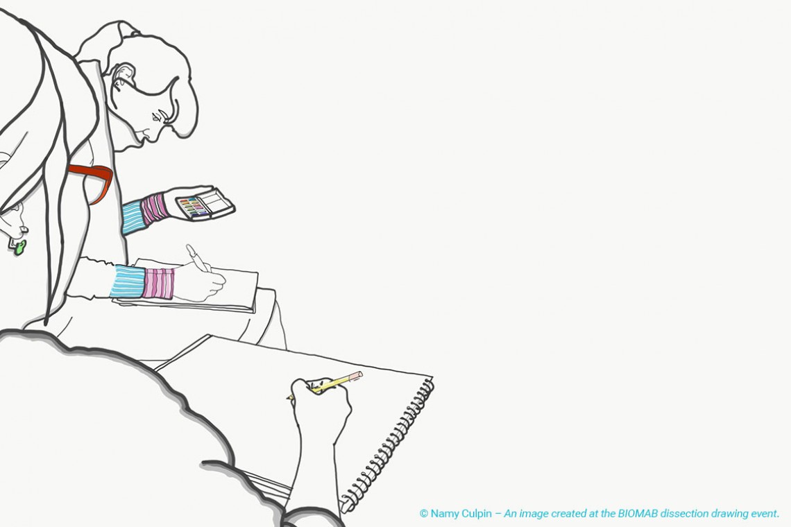
In both cases, I have found my experience to be exceedingly positive and informative. The demonstrators were very knowledgeable and explained the body structures and functions clearly and in detail. We were given opportunities to ask questions, take photographs and make sketches, which greatly enhanced my understanding of the subject. After the dissections, we were able to have free time to observe and draw other specimens at our leisure.
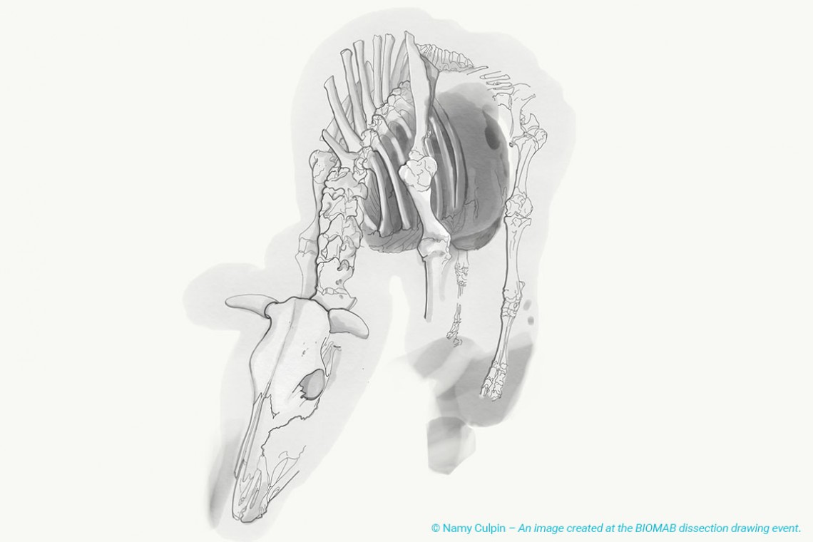
The dissection room at University of Dundee focused on human cadavers, while BIOMAB included both human and animal ones. I enjoyed learning about and drawing human and animal anatomy together, and I feel that this improved my general understanding of the anatomical structures.
In addition to the knowledge and skills I gained from these experiences, I also felt that I was in an environment of utmost respect and consideration for the body donors, as well as for my fellow peers and colleagues.
In my time spent studying medical art, I have had the privilege of dissecting cadavers to enhance my anatomical knowledge and apply it to art. I had dissected a variety of mammals preserved in formaldehyde prior to my graduate studies at University of Dundee.
I was especially inspired by my new experiences of dissecting Thiel-embalmed cadavers. University of Dundee allowed me access to cadavers that exhibited the colours and flexibilities of live humans. It was the quality of these cadavers that led to a shock factor throughout this semester. I had the feeling that these bodies were not deceased, just selflessly loaning their physical presence to us. Though I grew familiar with these cadavers, I was always still shocked when they were unveiled for a session of either dissecting or drawing.
I was also given the opportunity to experience a fresh/frozen dissection drawing day at BIOMAB in Antwerp, Belgium. These freshly deceased cadavers were dissected throughout the day for those attending, allowing me the time to focus solely on artwork as well as noticing the brilliant colour change within the limbs as the cadavers began to decompose throughout the eight-hour session.
As I proceed in my medical illustration career, I continue to appreciate and convey the memory of what each of these cadavers have taught me within my work. These bodies have left a lasting impression as I apply anatomy to art. I firmly believe that cadavers are an irreplaceable learning resource in this field.
The 6th ARS (Art Researches Science) Dissection Drawing Event took place on the 8th and 9th March 2013 in Ghent and Antwerp, Belgium. The event brought together over 60 practicing and student medical artists, illustrators, graphic designers, fine artists and anatomists for two days of observational drawing, dissection sessions and discussions from all over Europe.
There were attendees from numerous higher education courses and institutions such as the Medical Artists' Education Trust, London; Royal College of Art and University of the Arts MA Art & Science course, London; Haute école des arts du Rhin à Strasbourg; Royal Academy of Fine Arts in Antwerp; University of Antwerp, Faculty of Medicine and Health Sciences and the MSc Medical and Forensic Art courses, Dundee.
This year’s event promised to be different than in past years as the first day would be spent observing animal dissection and the second observing human dissection, allowing chance for comparative anatomy discussion.
The event began in the Historic city of Ghent where all the attendees had a chance to meet others participating in the two day event and talk about their own practice, study and interests in anticipation of the drawing days ahead.
The venue for the first day of the event was the Faculty of Veterinary Medicine at the University of Ghent. All attendees met for an introductory lecture and tour of the department and facilities by two of the trainers. There were introductory presentations by representatives of the Universities and Colleges that make up the ARS and I was there to represent the MSc Medical and Forensic Art courses at the University of Dundee, along with two MSc Medical Art graduates.
Everyone was then invited to witness the dissection of the abdominal cavity of a dog in the impressive observation-dissection room. People were allowed to draw, photograph or simply watch as one of the trainers began the dissection and were encouraged to ask as many questions as possible. Once the dissection was complete the group was split into two smaller groups. One group moved to the main dissection laboratory to have hands on dissection experience with animal specimens, ranging from horse and dog to pig, with the other group moving into the large animal anatomy museum for a chance to complete some observational drawing from preserved specimens.
In the dissection lab the anatomists continued to dissect and talk through the anatomy of the animals present, while giving the opportunity for people to dissect and sit and draw. The anatomy museum housed skeletons of giraffe, elephant and dolphin to name a few and also some pathological and fantastical specimens such as conjoined twin calves and two-headed piglets. There were also many corrosion casts of vessels and taxidermy specimens to view and draw.
The event dinner was held in a local restaurant and gave opportunity for all those present to talk about the day’s session and to share tips, tricks and knowledge.
The venue for the second day of the event was the Faculty of Medicine and Health Sciences at Antwerp University. Here in the dissection laboratory, anatomists two of the trainers and two anatomy PhD students dissected and explained in close detail an upper and lower arm (deep frozen and thawed), an upper and lower leg (deep frozen and thawed), a full brain, a heart with its coronary vessels and two lungs with their network of bronchi. Dissection of the arm and leg happened simultaneously allowing the viewer to move back and forth between tables and pick and choose what was most interesting to draw.
There were moments in which dissection was temporarily halted to allow the artists to sketch and create artwork from the specimens, while other participants moved in to dissect and discuss the anatomy in more detail with the demonstrators. There were also opportunities to draw from preserved specimens and there were medical models and textbooks on hand to aid in the understanding of some of the anatomy.
The day concluded with an exhibition of the artwork produced over the two days and gave an opportunity to discuss technique, drawing style, materials used and provided everyone present with a chance to ‘show off’ their artwork and learn from each other.
The Drawing Dissection event ended with a meal at a German beer house in the fantastic centre of Antwerp and a lively discussion in a pub overlooking the cathedral at night. The event was a wonderful opportunity to meet other medical artists and those who use images of anatomy either as inspiration or in their artwork. For me, being able to see, dissect and illustrate deep frozen and thawed human specimens was the highlight of the event.
As this is a growing international art event, an overview exhibition and publication of the large amount of artwork that has been produced and will be organised and showcased in 2014.